Continuing our essay series on our bodies’ electric current, primarily through Frances Ashcroft’s The Spark of Life: Electricity in the Human Body, and Sally Adee’s We Are Electric: Inside the 200-Year Hunt for Our Bodies’ Electric Code, and What the Future Holds, this essay explores the human heart, and how our senses work.
As Ashcroft writes:
In essence, your heart is a pump that is controlled by electricity. Blood enters via the upper chambers (the atria), which contract first and force blood into the much larger lower chambers (the ventricles). The ventricles contract in synchrony about half a second later, the right ventricle pumping blood to the lungs and the left ventricle sending it around the body. Non-return valves lie between the upper and lower chambers of the heart so that the blood only flows in one direction; from the atria to the ventricles. Similarly, non-return valves guard the exits from the ventricles into the great vessels. If these valves leak, as can happen with age, then blood is pumped less efficiently, so that the body receives less oxygen and you feel constantly tired. The chambers on the right and left side of the heart are physically separate which ensures that oxygen-rich blood coming from the lungs is not mixed with oxygen-depleted blood returning from the tissues. However, because heart cells are wired together, they contract in synchrony, so that the heart beats as a single organ … The time lag associated with electrical transmission ensures that the electrical signals reach the upper chambers before the lower ones, so that first the atria are triggered to contract, and then the ventricles. The timing of this spread of excitation is crucial for the ability of the heart to serve as a pump. If it is disrupted, the heart no longer beats regularly and its capacity to pump blood is compromised … As in the case of nerve cells, ion channels are responsible for the electrical impulses of heart cells. However, many more types of channel are involved in shaping the action potential of the heart. It is initiated by the opening of sodium channels. These channels are similar, but not identical to those of nerve cells, which explains why fatal poisons like that of the puffer fish block electrical impulses in the nerves, but not the heart … Opening of the sodium channel pores is quickly followed by the opening of calcium channels, which enables calcium ions to flood into the cell, where they trigger the release of stored calcium and thereby contraction. Calcium channels are not just important for letting in the calcium ions that trigger the release of stored calcium. The fact that they close (inactivate) only slowly at positive membrane potentials helps prolong the cardiac action potential, thereby providing more time for the heart to contract. The action potential of a ventricular cell is about half a second long, almost 500 times longer than that of a nerve cell … The end of the cardiac action potential is produced by opening of potassium channels, and the resulting efflux of potassium ions returns the voltage gradient across the membrane to its resting value. As a consequence, the calcium channels shut, preventing calcium influx, so that the heart relaxes.
Ashcroft then describes how our human senses work using our body’s electrical impulses. She starts with the eye:
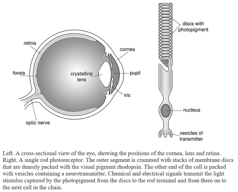
Pleasure, pain, indeed the evolutionary success of any organism, ourselves included, depends on our ability to perceive the world around us: to see, hear, smell, taste and touch it. Our sense organs convert the myriad signals that constantly bombard us in multiple modalities into a single form that the brain can interpret – the electrical energy encoded in our nerve impulses. And in all cases ion channels are needed to transduce sensory information into that electrical signal. Ion channels are truly the doors of perception as everything we sense is detected, transmitted or processed by them … At one level, our eyes operate like a simple camera. They have a clear cornea and a crystalline lens that work together to focus light rays onto a layer of photosensitive cells known as the retina at the back of the eye. They possess an iris that continuously adjusts the amount of light entering the eye. And they have a protective lens cap – the eyelid – that can shut the light out completely when necessary. Unlike most cameras, however, our eyes have a brain attached that processes and interprets the images projected on the retina. Some processing also takes place in the retina itself. Every second, our eyes handle thousands of images, transforming light signals into upside-down images on the retina, and converting these into nerve impulses that are sent on to the brain for processing. The transparent outer layer of your eye, the cornea, is responsible for about two-thirds of the focusing power of the eye: the remainder is the province of the lens, which is suspended behind the pupil by thousands of fine ligaments. The cornea has a fixed focus, but that of the lens can change, for muscles attached to its edge pull it thicker or thinner as you focus on near and far objects, respectively. As we grow older the elasticity of the lens decreases, making it harder to change its focus, which is why most people over fifty need reading glasses. The pupil is the aperture through which light passes. It appears black because no light returns through it. The iris – the coloured bit of your eye – contains muscles that adjust the size of the pupil to the intensity of the ambient illumination, dilating it in dim light and shrinking it to a pinpoint in very bright light. The size of the pupil even signals feeling, for it expands in response to fear, pain or if you see something of interest – someone you love, perhaps.
Our ability to see begins with this transduction of light into an electrical signal, in which ion channels play an intimate role … [P]hotoreceptors employ an intracellular messenger service to link the light-induced conformational change in the photopigment to transmitter release. This consists of a chemical known as cyclic GMP, which relays information from the intracellular discs to the surface membrane of the cell. Here, the chemical signal is converted into a much faster electrical one that is rapidly conducted to the transmitter release sites. At the heart of this complex signalling cascade is a special kind of ion channel that opens when it binds cyclic GMP. In the dark, cyclic GMP levels in the rods and cones are high, so that the cyclic GMP-gated channel is held open. Sodium ions flooding in through the channel pore produce a positive swing in the membrane potential that spreads over the surface membrane to the other end of the rod or cone cell. There, it stimulates calcium channels to open, enabling calcium ions to enter the cell and trigger the release of a transmitter that stimulates the next cell in the chain and thereby tells the brain that it is dark. Light-induced changes in the visual pigments activate a signalling cascade that leads to the destruction of cyclic GMP. As a consequence, the cyclic GMP-gated channel closes, switching off transmitter release and signalling ‘Light!’ The exquisite sensitivity of our vision derives from this complicated chain reaction, which constitutes a powerful amplification system.
Ashcroft then describes how the ears operate:
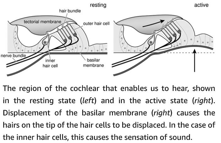
The outer and inner chambers of the ear, showing how displacement of the ear drum is transmitted via the three ear bones (malleus, incus and stapes) to the fluid-filled cochlea where the hair cells transduce sound vibrations into electrical impulses. An elegant description of how we hear is given by Aldous Huxley in his novel Point Counter Point. ‘Pongileoni’s bowing and the scraping of the anonymous fiddlers had shaken the air in the great hall, had set the glass of the windows looking onto it vibrating; and this in turn had shaken the air in Lord Edward’s apartment on the further side. The shaking air rattled Lord Edwards’ membrana tympani; the interlocked malleus, incus and stirrup bones were set in motion so as to agitate the membrane of the oval window and raise an infinitesimal storm in the fluid of the labyrinth. The hairy endings of the auditory nerve shuddered like weeds in a rough sea; a vast number of obscure miracles were performed in the brain, and Lord Edwards ecstatically whispered “Bach!”’ As Huxley relates, sounds are simply pressure waves in the air that radiate outward from a sound source, much like the ripples in a pond. These are collected and filtered by our ears and funnelled towards the eardrum, which vibrates in response. In turn, this moves three delicate interlocking bones, the malleus (‘hammer’), incus (‘anvil’) and stapes (‘stirrup’), which are among the smallest bones in our body and no bigger than the size of a single letter of this print. These relay the vibrations to another membrane, the oval window. At this point the sound waves pass from air to the fluid-filled canals of the inner ear, where sensory cells convert the sound waves into electrical impulses. These are then forwarded via the auditory nerves to the brain, where they are interpreted.
Sound waves induce oscillations in the cochlear fluid on either side of the basilar membrane, causing it to vibrate. The tiny movement of the basilar membrane is transferred to the hair cells, causing their stereocilia to be displaced backwards and forwards, so producing a mechanical deflection that opens specialized ion channels. These mechanically gated ion channels are at the heart of hearing for it is they that convert sound waves into electricity – or more precisely mechanical energy into electrical energy. The stereocilia the hair cells bear are organized in rows of decreasing height and are connected to one another at their tips by a tiny thin stiff rod known as a ‘tip link’. One end of the tip link is also connected to the mechanosensitive channels that sit at the tip of the stereocilia. As the basilar membrane moves up and down, the tip links are stretched or compressed, pulling open, or forcing shut, the ion channels, respectively. When they open, positively charged ions rush into the cell and alter the voltage gradient across the hair cell membrane. This electrical change has different effects depending on whether the cell is an inner or an outer hair cell. The change in voltage across the inner hair cell membrane produced by sound of a specific frequency triggers the release of a chemical transmitter. This stimulates impulses in the terminals of the auditory nerve and so sends signals to your brain. I’ve not forgotten the time I visited Jonathan Ashmore’s laboratory at University College London and looking down the microscope was amazed to see a tiny hair cell bopping away to ‘Rock around the Clock’ … The deterioration in hearing we all experience with age is caused because our hair cells naturally die off with time and once gone they are lost forever. Loud noises destroy our hearing even faster. The rock musician Pete Townshend7 instantly lost the hearing in one ear in a famous incident in which a staged explosion was far louder than expected. Thousands of troops fighting in Iraq and Afghanistan returned home with permanent hearing loss, mostly caused by roadside bombs. The thunder of warfare, the deafening sound of a pop concert, the roar of jets and loud machinery – all exact a heavy toll. This is because our outer hair cells are very vulnerable and can be irretrievably damaged by loud noises.
Ashcroft then describes how our taste buds work:
Taste cells are not nerve cells but a specialized kind of epithelial cell (the cells that line the gut, mouth and nasal passages). They are very short-lived, being continually replaced every couple of weeks, and they are packed together in barrel-shaped taste buds. Humans have about 10,000 taste buds distributed over the surface of the tongue, each containing 50 to 100 taste cells.9 Each taste cell sends a long finger-like process, tipped with fine hairs that bear the taste receptors, up to the opening of the taste bud on the surface of the tongue where stimuli are received. The other end of the taste cell contacts the sensory nerve. We can discriminate five basic tastes – sweet, salt, sour, bitter and savoury (umami). All the many different flavours we taste, however, are really smelt, for these two senses work in combination. This explains why your sense of taste seems impaired when you have a head cold and your nose is blocked. When you eat something, chemicals contained in the food dissolve in your saliva. This enables them to bind to the receptors at the tip of the taste cells, and so trigger a cascade of events that ultimately releases a chemical transmitter from the base of the taste cell. In turn, this excites the sensory nerve, and nerve impulses are then transmitted to the brain where the information is decoded, processed and tastes are identified. Different tastes arise because different types of receptor are stimulated. Two tastes – salt and sour – are directly detected by ion channels sensitive to the ions involved, which are respectively sodium ions and hydrogen ions (protons). Salty tastes are mediated by the epithelial sodium channel (ENaC) we met in the previous chapter. Several kinds of ion channel that are sensitive to protons detect sour tastes. The carbon dioxide in fizzy drinks and champagne is also detected by sour taste receptors because it yields protons when dissolved in water. Interestingly, some soda-water manufacturers recognized this long before science showed it to be true – sauerwasser10 and similar seltzers are named for their sour, slightly acidic taste. Umami, from the Japanese word umai, meaning ‘delicious’, describes the savoury taste of food containing monosodium glutamate. Some of the receptors that detect glutamate are also ion channels. Sweet and bitter substances do not activate ion channels directly. Instead they bind to specific receptors, so setting in train a cascade of biochemical events that eventually leads to the opening of a specialized ion channel (known as TRPM5) that is common to both pathways. The ability to discriminate between sweet and bitter substances arises because the two types of receptors are found in different populations of taste cells, which signal separately to the brain. Thus whether something tastes sweet or bitter is decided by the brain. We have over twenty different receptors for bitter taste, but only one for sweet taste, reflecting the evolutionary drive to identify bitter-tasting substances, which are often poisonous.
And then our sense of smell:
The cells that detect smells lie high up in the nose, almost seven centimetres away from the nostril. These are the olfactory neurones, which send processes to the olfactory epithelium in the nose. Each nerve process terminates in a small bunch of olfactory cilia, fine hair-like processes that project up into the viscous mucous layer that covers the moist surface of the inner nose and greatly increase the membrane surface area available for odorant detection. Odorant receptors lie embedded in the surface of the cilia, ready to capture smells borne on the air you breathe. Humans have around 350 distinct types of olfactory receptor proteins,11 although each olfactory neurone carries only a single kind. But we can detect far more than 350 aromas: most people can distinguish many thousands of substances, often in tiny amounts. In the same way that the letters of the alphabet can be used to construct a vast vocabulary, so the different combinations of odorant receptors produce a cornucopia of pure odours. One reason for our supposed poor sense of smell is that we walk around with our noses high in the air, while scents are at their strongest close to the ground and quickly dissipated by air currents at higher levels, as can easily be seen by watching how a tracker dog follows a trail. Precisely how odorants stimulate their receptors is still unclear, but it appears to be due to the different sizes and shapes of the odorant molecules. One idea is that they bind to the receptor in a lock-and-key fashion. Just as your right glove will only fit your right hand, so right-handed molecules will only bind to right-handed receptors; this explains why orange and lemon (which are left- and right-handed versions of limonene) smell different. Binding of an odorant to its receptor triggers a cascade of events in the neurone that leads to opening of a specific kind of ion channel – related to, but different from, those in the rods and cones – so giving rise to a current that in turn sets up a stream of action potentials in the olfactory neurone itself. These impulses pass along the olfactory nerve to a region of the brain known as the olfactory bulb, where they hand their signals on to other nerve cells in deeper regions of the brain … Like most sensations, if you are exposed to a constant smell you gradually become accustomed to it and eventually no longer perceive it. Most people fail to notice their own body odour, or even the perfume they are wearing after a while.
And finally, Ashcroft describes how our sense of touch works:
From the caress of a loved one, to the feel of the wind on our cheek or the crush of a bear-hug, touch plays an important part in all our lives. Sense organs in our skin respond to such mechanical forces with an electrical change, triggering nerve impulses that relay information back to the spinal cord and brain. Like other sensory nerves, impulse frequency is graded according to the stimulus strength, with lighter touches evoking fewer impulses than stronger pressures. Touch receptors also adapt to continuous stimulation, which explains why we do not notice the pressure of the clothes we wear. Exactly how mechanical energy is transformed into electrical energy remains a puzzle, but it is clear that mechanically sensitive ion channels are somehow involved. Recent studies suggest that these channels are attached to the extracellular surface of the cell by a gating tether, in an arrangement similar to that found in the hair cells of the ear. Pressure on the cell membrane is thought to tug on the tether, distorting the structure of the channel so that it opens. The more the membrane is deformed, the more channels are likely to be activated and the greater the excitation of the nerve. Sometimes nerve endings sensitive to mechanical force are packaged into specialized structures that enhance their ability to detect changes in pressure or vibrations such as those that arise when you stroke your fingers across a rough surface. However, the end result is the same: a mechanical stimulus elicits an increase in action potential frequency in the sensory nerve. Our skin not only contains receptors sensitive to pressure, but also to temperature and painful stimuli … [T]he reason chilli peppers taste so hot is that they open the same ion channel as high temperature, and because the brain cannot tell the difference between the two stimuli it interprets them both as heat. These channels are not just found in the tongue, they are also present in the skin of your fingertips, face and other sensitive parts of the body – as unfortunate men who have been chopping chilli very quickly find out if they forget to wash their hands before visiting the lavatory … Just as chilli stimulates hot receptors, so other chemicals interact with receptors that sense cold, fooling the body into thinking the substance is cool. The minty, fresh taste of menthol, found in peppermint oil, arises from the fact it activates an ion channel that detects cold temperatures … Pain can be extremely useful – it is a valuable alarm system that signals danger. It tells us that the pan is hot; that our toes are in the fire; that straining too hard will tear our muscles; that we have an infection or a wound. Without it you may be burnt, develop suppurating sores, or walk around with broken limbs, causing further damage. Pain also reminds us to allow damaged parts of the body to heal. All pain comes from the brain. It is our brain that receives messages from nerve fibres, telling us we have stubbed our toe, and many brain areas are involved in our experience of pain; they tell us where the pain is, how much it hurts, and what kind of pain it is – sharp, burning or just a dull ache. Our perception of pain is also highly variable. Even if the input signal from our sensory nerve endings is identical, the way the signals are processed is powerfully influenced by our attention, mood and expectation and this can dramatically alter the pain we experience. Our emotions can make a placebo pill an effective painkiller even though it contains no active ingredient, and, conversely, fear of pain can sharpen its impact … We are preprogrammed to respond most strongly to changes in our environment and cease to pay attention if nothing new happens, a phenomenon that has a clear evolutionary advantage.
In the next essay in this series, we’ll explore the human brain.


